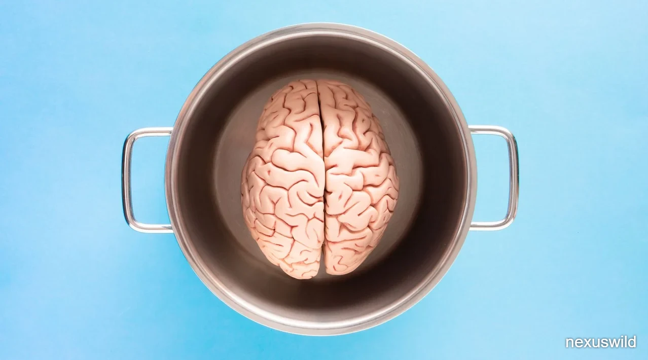

The brain does not switch off during sleep. In the deepest stages, slow electrical waves sweep through the cortex, and rhythmic pulses of cerebrospinal fluid move through the spaces around the brain, carrying waste products away. MIT researchers have now found that this fluid movement is markedly weaker in older adults, and that the decline tracks changes in both brain-wave activity and the blood vessels that supply the brain.
The study, published in Nature Aging, compared adults aged 18 to 39 with adults aged 60 to 84. Participants were monitored with a combination of electroencephalography, functional magnetic resonance imaging and a specialised MRI method that tracks cerebrospinal fluid motion. Many fell asleep inside the scanner, allowing the team to compare wakefulness and non-REM sleep in the same person.
The result was a clear age difference. In younger volunteers, the slow waves of deep sleep were accompanied by larger waves of cerebrospinal fluid. In older volunteers, those fluid waves were substantially smaller on average. The researchers report that the difference cannot be explained simply by older adults spending less time asleep.
The team then looked for explanations in the vascular system. In separate experiments conducted while participants were awake, they measured how well blood vessels responded to increased neural activity and to raised carbon dioxide levels. Older volunteers showed weaker responses in both tests. A statistical model suggested that these vascular changes account for much, but not all, of the age difference in fluid movement.
The finding does not show that weaker cleansing causes dementia, and none of the volunteers had been diagnosed with a neurodegenerative disorder. It does, however, add a physiological detail to a growing picture of how sleep, blood flow and brain health interact during aging. The authors hope to follow participants over time and to understand why some older adults retain fluid waves similar to those seen in healthy younger people.
The question: does cleanup fade with age?
The starting point for the study was a discovery the same laboratory made in 2019. Working with human participants, the MIT team found that slow electrical waves during non-REM sleep are coupled to rhythmic movements of cerebrospinal fluid. Those waves appear to help drive fluid through the spaces surrounding the brain, a process linked to the removal of metabolic waste.
That earlier work raised an obvious follow-up. Slow brain waves are known to change with age, typically becoming less pronounced in older adults. If the waves help generate the fluid pulses, might the pulses also weaken as people get older?
The scientific literature before this study was not consistent. Some investigations had reported that cerebrospinal fluid movement declined with age, while others reported increases. According to the MIT researchers, a key limitation was that many of those studies did not monitor fluid movement during sleep, when the brain's cleaning activity is most relevant.
The new study was designed to address that gap directly. Rather than inferring fluid dynamics from structural scans or daytime measurements, the team recorded what happened as participants moved from wakefulness into sleep. That allowed them to connect three signals in the same person: the brain's electrical rhythm, the flow of blood through the brain, and the motion of cerebrospinal fluid.
The question has implications beyond normal aging. Sleep disruption has been associated in previous research with higher risk of Alzheimer's disease and other forms of dementia. If a measurable change in fluid movement accompanies aging, it may eventually help researchers identify people whose brains are changing in ways that merit closer attention.
How the team observed fluid and sleep
No single imaging method can capture all of the processes the researchers wanted to measure, so they combined several. Electroencephalography, or EEG, recorded the brain's electrical activity and allowed the team to identify slow waves and stages of sleep. Functional MRI tracked changes in cerebral blood flow, showing how blood vessels responded to neural activity. A specialised MRI technique developed in Laura Lewis's laboratory measured the movement of cerebrospinal fluid.
Bringing these methods together inside a scanner is technically demanding. MRI is noisy and constraining, and sleeping inside the instrument is not the same as sleeping at home. The researchers scheduled sessions late at night to increase the likelihood that participants would become drowsy or fall asleep during the measurements. Each session lasted approximately two hours.
The design gave the team a within-person comparison. For participants who fell asleep, the researchers could contrast periods of wakefulness with periods of non-REM sleep, rather than comparing different groups of people under different conditions. That matters because fluid movement varies from person to person, and a within-person comparison reduces the influence of those baseline differences.
The analysis focused on the relationship between slow electrical waves and fluid pulses. In the younger group, the appearance of stronger slow waves during non-REM sleep was accompanied by larger cerebrospinal fluid waves. In the older group, that coupling was weaker and the average fluid movement was substantially reduced.
The authors were careful to note that a smaller fluid wave is not the same as a blocked cleaning system. The measurement captures the rhythmic component of fluid motion associated with sleep. It does not measure every route by which the brain clears waste, nor does it establish that the total amount of clearance fell by a given percentage.
The volunteers and the scanning session
The study included two age groups. Younger participants were between 18 and 39 years old, while older participants were between 60 and 84. The age ranges were broad enough to capture meaningful differences in brain physiology but narrow enough to keep each group reasonably coherent.
Each volunteer took part in a testing session of roughly two hours, beginning late at night. The timing was a practical choice: the team wanted participants to be sleepy during the scan, so that they could observe the transition into non-REM sleep under controlled conditions. Sleep in a scanner is often lighter and more fragmented than sleep in a bedroom, a limitation the researchers would need to consider when interpreting their results.
While in the scanner, participants were monitored with EEG, fMRI and the cerebrospinal-fluid imaging sequence. The combination allowed the team to line up the timing of electrical events with changes in blood flow and fluid motion. That temporal alignment was central to the analysis, because the question was not only how much fluid moved but when it moved relative to the brain's electrical cycles.
In a separate set of experiments, participants remained fully awake while the researchers tested blood-vessel function. Those experiments did not depend on sleeping inside the scanner and were designed to isolate a possible mechanism for the age difference seen during sleep.
No participant in the study had been diagnosed with a neurodegenerative disorder. That criterion is important for interpretation: the observed differences belong to aging brains without a clinical diagnosis, rather than to brains already affected by dementia. Some older participants did show signs of gray matter atrophy on their scans, which the team examined separately.
What changed during deep sleep
The central observation was straightforward. In younger adults, entering non-REM sleep brought stronger slow brain waves and larger cerebrospinal fluid waves. In older adults, the fluid waves were much smaller on average.
The researchers emphasised that the reduction was not simply a consequence of older adults sleeping less or spending less time in deep sleep. Because the team compared periods of wakefulness and sleep within the same participants, they could account for time asleep. The difference remained after that consideration.
The result fits with a broader pattern of age-related change in sleep architecture. Slow-wave sleep tends to become lighter and less consolidated with age, and the electrical waves that characterize it can lose amplitude. The MIT work extends that observation by showing that the fluid dynamics linked to those waves change as well.
The team did not report that every older participant showed the same decline. Average fluid waves were smaller in the older group, but individual outcomes varied widely. That variation became one of the most intriguing aspects of the study, because it suggests that some brains preserve a physiological feature that others lose.
For now, the finding is observational. It establishes an association between age, sleep-wave activity and fluid movement. Whether the change in fluid movement has consequences for cognition or for the accumulation of proteins associated with neurodegenerative disease is a question the researchers plan to investigate in longitudinal follow-up.
Testing the blood-vessel explanation
Having found an age difference in fluid waves, the team turned to the question of why it occurs. One candidate was neurovascular coupling, the process by which active brain regions receive additional blood. When neurons become more active, nearby blood vessels normally widen to deliver oxygen and nutrients. If that response weakens with age, the rhythmic changes in blood volume that help drive cerebrospinal fluid could also diminish.
To test the idea, the researchers showed participants a flickering checkerboard pattern while measuring brain activity and blood flow with fMRI. A rapidly changing visual pattern reliably stimulates visual cortex, which should trigger a local increase in blood supply. In many older participants, electrical activity rose as expected, but the accompanying rise in blood flow was much smaller.
That result points to a mismatch between neural demand and vascular response. The brain cells responded to the stimulus, but the vessels feeding them did not fully compensate. Such a mismatch could alter the pressure and volume changes that accompany slow-wave sleep, and therefore the fluid waves coupled to them.
The experiment was conducted while participants were awake, because a controlled visual stimulus is easier to interpret than the fluctuating activity of sleep. The assumption is that the vascular responsiveness measured in this way reflects a property of the brain's circulation that also operates during sleep. That is a reasonable inference, but it remains an inference rather than a direct measurement of the same process in both states.
The authors also note that vascular health is influenced by many factors, including lifestyle and medical history. Their experiments measured two specific responses, not the full range of changes that occur in aging blood vessels.
Breath-holding and carbon dioxide response
The second vascular test used a different challenge. Participants were asked to hold their breath while researchers monitored cerebral blood flow. Holding the breath causes carbon dioxide to accumulate in the bloodstream, which normally prompts blood vessels in the brain to widen and allow more blood to pass through.
Once again, the older group showed a weaker response. On average, their cerebral blood flow increased less than that of younger volunteers. Together with the checkerboard experiment, this result suggests that the aging brain's circulation is less flexible in responding to both neural activity and chemical signals.
The breath-hold test is a standard way to probe vascular reactivity because carbon dioxide is a potent dilator of cerebral blood vessels. A reduced response can indicate that vessels are stiffer, that the signalling pathways controlling them are less effective, or that the maximum achievable flow is lower. The MIT study did not distinguish among those possibilities.
What the two experiments do establish is that older participants, as a group, showed weaker vascular responses in two quite different situations. That consistency strengthens the case that vascular changes contribute to the smaller sleep-related fluid waves observed in the same participants.
The researchers used a statistical model to estimate how much of the fluid-wave difference could be attributed to the two vascular measures. The model accounted for a substantial portion of the decline, but not all of it. In other words, vascular changes appear to be important without being a complete explanation.
What the statistical model could and could not explain
The modelling step was designed to move the study beyond simple correlation. The team asked whether the measured differences in neurovascular coupling and carbon-dioxide responsiveness could account for the difference in cerebrospinal fluid waves between the age groups.
The answer was partial. Those two factors explained much of the observed decline, which supports the idea that blood-vessel function matters. But a residual difference remained, meaning other age-related changes must also be involved. The authors did not claim to have identified those additional factors.
That caution is deliberate. Aging affects the brain in many ways at once: tissue structure, vascular health, sleep architecture, cellular metabolism and the chemical environment around neurons. Separating their individual contributions is difficult, particularly in a study of this size and duration.
Sydney Bailes, one of the study's lead authors, has noted that the complexity of aging makes it hard to disentangle the relevant mechanisms. Even with detailed measurements, factors such as overall vascular health and lifestyle may influence the results in ways that the current model cannot capture. Measuring all of them would be required to fully break down the reasons one person's fluid flow is more or less affected than another's.
The team's next step is to follow participants over time. Longitudinal data could show whether weaker fluid waves predict later cognitive change, and whether the vascular measures collected in this study help explain that relationship. Such work would be needed before the findings could be used to identify people at elevated risk.
Gray matter atrophy and early warning signs
Although no participant had a diagnosed neurodegenerative condition, brain scans revealed that some older adults showed signs of gray matter atrophy. Gray matter contains large numbers of nerve cells, and loss of tissue in these regions is a feature of several age-related brain disorders.
The researchers found that greater atrophy was associated with larger reductions in cerebrospinal fluid movement. That association is biologically plausible: changes in brain tissue and its supporting circulation could both contribute to weaker fluid dynamics. But the study's design cannot establish direction. It does not show that reduced fluid flow causes tissue loss, nor that tissue loss causes reduced flow.
The authors treat the association as a reason for further investigation rather than a clinical finding. They want to know whether cerebrospinal fluid waves measured during sleep could eventually serve as an early indicator of neurological decline. Testing that idea would require following participants for years and comparing the imaging measures with cognitive outcomes and diagnoses.
The result also connects the study to a longer research tradition. Sleep disturbance has been linked to Alzheimer's disease risk in epidemiological work, and cerebrospinal fluid is the medium that carries several proteins implicated in that disease. Understanding how sleep-driven fluid motion changes with age could eventually clarify one pathway connecting the two.
For now, the team stresses that the finding applies to group averages and associations. It is not a diagnostic test, and it cannot be used to tell an individual that their brain is clearing waste poorly.
The exception: older adults with youthful waves
One of the most striking features of the data was its variability. While older participants had smaller fluid waves on average, some retained movements within the range measured in healthy younger adults. The researchers describe this subgroup as particularly intriguing.
Laura Lewis has pointed out that people age very differently. Some maintain strong cognitive function into later life, while others develop decline. If researchers can identify what distinguishes those trajectories, they may be able to predict future outcomes more accurately. The preservation of sleep-related fluid waves could be one such marker, although the current study does not show that it is.
The observation raises several possibilities. The older adults with stronger waves may have better vascular health, more preserved sleep architecture, genetic factors that protect brain tissue, or lifestyle characteristics that support brain function. The study was not designed to test these explanations, and the authors have not reported differences in lifestyle or medical history between the subgroups.
Identifying the factors involved would require a larger and more diverse sample, with detailed information about cardiovascular health, medication use, sleep habits and other variables. It would also require long-term follow-up to determine whether preserved fluid waves correspond to better cognitive outcomes.
The finding is nevertheless important because it shows that the age-related change is not inevitable in every individual. That opens the door to questions about whether the processes underlying it can be supported or restored, rather than simply accepted as a consequence of growing older.
Pink noise and the search for interventions
The question of whether brain cleansing can be strengthened is already being explored in the same laboratory. A separate recent study from Lewis's group found that brief bursts of pink noise during sleep could increase cerebrospinal fluid movement in younger adults. Pink noise contains a broad range of frequencies with relatively more energy at lower frequencies than white noise.
The approach is non-invasive. Instead of delivering a drug or stimulating the brain electrically, researchers play carefully timed sounds during sleep to reinforce the slow waves that drive fluid movement. Earlier work by other groups has explored related forms of acoustic stimulation as a way to enhance slow-wave sleep and memory.
The important limitation is that the pink-noise experiment was conducted in younger adults. Researchers have not yet tested whether the same approach can increase fluid movement in older people, whose brain waves and vascular responses differ. It is therefore unknown whether the method would help the group most affected by the decline described in the new study.
Other possible interventions remain speculative. Exercise has been studied in relation to the brain's waste-clearance systems, and some research suggests physical activity may support processes linked to what is often called the glymphatic system. Whether such measures would change the specific fluid waves measured by the MIT team is not established.
The authors are interested in testing interventions, but they present that work as a future direction. The immediate contribution of the current study is a better description of the problem, including which physiological systems appear to be involved and which do not fully explain it.
What comes next for the research
The study was funded by the National Institutes of Health, the Simons Collaboration on Plasticity in the Aging Brain, a McKnight Scholar Award, a Pew Biomedical Scholar Award, an MIT EECS Transformative Research Award, the Picower Institute Neurological Disorder Discovery Research Fund, a Sloan Fellowship and a Department of Defense National Defense Science and Engineering Graduate Fellowship Program.
The paper lists Sydney M. Bailes and Stephanie D. Williams as lead authors and Laura D. Lewis as senior author. The full author list includes collaborators whose contributions ranged from imaging analysis to clinical and anatomical expertise. The research was published in Nature Aging under the title "The aging human brain exhibits reduced hemodynamic and cerebrospinal fluid flow waves during sleep."
The team's stated priorities are clear. They want to follow participants to see who develops cognitive problems and whether baseline fluid waves help predict that outcome. They want to understand what distinguishes older adults whose waves resemble those of younger people. And they want to test whether interventions can strengthen the process.
Until that work is done, the result should be read as a physiological observation with potential implications rather than a clinical tool. It adds detail to the ways aging changes the sleeping brain, and it gives researchers a measurable phenomenon to investigate in relation to dementia risk. The researchers themselves describe the next steps as an effort to understand successful aging, not merely to catalogue decline.
Nexuswild welcomes factual corrections. Email contact@nexuswild.com with evidence and the article URL.